미국 NIH (보건원)에서 발간한 사람 시신의 조직(뇌, 신장, 간 등) 에서 검출된 미세플라스틱에 대한 전세계 첫 사례를 공유드립니다.
논문의 결론에서는
- 뇌는, 간 또는 신장 시료보다 MNP(미세 및 나노플라스틱) 농도가 더 높았습니다.
- 모든 장기는 2016년에서 2024년 사이에 상당한 증가를 보였습니다.
본 연구결과는 알츠하이머 또는 치매와 같은 뇌질환과 MNP간의 연관성 연구에 중요한 연구자료의 첫 사례로 판단됩니다.
이러한 실제 인체 시료 중에 미세플라스틱에 대한 연구 필요성이 크므로 한국에서도 조기에 주요 질환군 시료를 대상으로 연구를 시작하길 희망합니다.^^
한국분석과학연구소는 본 논문에서 사용된 Py-GC-MS (TED-GC-MS) 및 FTIR, Raman장비를 확보하여 현재 국내 의학병원과 함께 임상시험심사위원회(IRB) 심사를 통과한 후 의뢰된 생체 시료(혈액 및 양수/조직 등)를 이용하여 분석법을 확립하였고, 그 결과 미세플라스틱 분석결과를 제공하고 공동논문 작업을 진행하고 있습니다.
관련 공동연구를 희망하시는 연구진께서는 언제든지 연락 바랍니다.
info@kiast.co.kr, 02-6951-1116


Bioaccumulation of Microplastics in Decedent Human Brains Assessed by Pyrolysis Gas Chromatography-Mass Spectrometry
Py-GC-MS법을 통해 평가한 사망자 뇌의 미세플라스틱 생물 축적
Abstract 초록
Rising global concentrations of environmental micro- and nanoplastics (MNPs) drive concerns for human exposure and health outcomes. Applying pyrolysis gas chromatography-mass spectrometry (Py-GC/MS) methods to isolate and quantify MNPs from human samples, we compared MNP accumulation in kidneys, livers, and brains. Autopsy samples from the Office of the Medical Investigator in Albuquerque, NM, collected in 2016 and in 2024, were digested for Py-GC/MS analysis of 12 polymers. Brains exhibited higher concentrations of MNPs than liver or kidney samples. All organs exhibited significant increases from 2016 to 2024. Polyethylene was the predominant polymer; the relative proportion of polyethylene MNPs was greater in brain samples than in liver or kidney. Transmission electron microscopy verified the nanoscale nature of isolated particles, which largely appeared to be aged, shard-like plastics remnants across a wide range of sizes. Results demonstrate that MNPs are selectively accumulated into the human #brain and concentrations are rising over time.
환경 미세 및 나노 플라스틱(MNP)의 전 세계적 농도 증가는 인간 노출 및 건강 결과에 대한 우려를 불러일으킵니다. 열분해 가스 크로마토그래피-질량 분석법(Py-GC/MS) 방법을 적용하여 인간 샘플에서 MNP를 분리하고 정량화하여 신장, 간 및 뇌에서 MNP 축적을 비교했습니다.
2016년과 2024년에 수집된 뉴멕시코주 앨버커키의 의학 조사관실에서 부검 샘플을 소화하여 12가지 중합체의 Py-GC/MS 분석을 수행했습니다.
뇌는 간 또는 신장 샘플보다 MNP 농도가 더 높았습니다.
모든 장기는 2016년에서 2024년 사이에 상당한 증가를 보였습니다.
폴리에틸렌이 가장 우세한 중합체였습니다. 폴리에틸렌 MNP의 상대적 비율은 간 또는 신장보다 뇌 샘플에서 더 컸습니다. 투과 전자 현미경(TEM)은 분리된 입자의 나노 스케일 특성을 확인했으며, 이는 광범위한 크기에 걸쳐 오래된 파편과 같은 플라스틱 잔재로 나타났습니다.
연구 결과에 따르면 MNP는 인간의 뇌에 선택적으로 축적되며 농도는 시간이 지남에 따라 증가하는 것으로 나타났습니다.
Keywords: #Polymer #neuronal #autopsy #liver #kidney
#nanoplastics #폴리머 #신경 #부검 #간 #신장
#나노플라스틱
The ubiquitous presence of plastics, especially polymer-derived particulates ranging from 500 micrometers in diameter down to 1 nanometer, defined as micro- and nanoplastics (MNP), is a defining feature of the Anthropocene epoch1. The extent to which microplastics cause harm or toxicity is unclear, although recent studies associated MNP presence in carotid atheromas with increased inflammation and risk of future adverse cardiovascular events2,3. In controlled exposure studies, MNPs clearly enhance or drive toxic outcomes4–6. The mantra of the field of toxicology – “dose makes the poison” (Paracelsus) – renders such discoveries as easily anticipated; what is not clearly understood is the internal dose in humans.
To date, several studies have utilized visualization and spectroscopic methods to identify and count particulates in organs such as the lungs, intestine7, and placenta8. These methods are often limited to larger (>1–5μm) particulates, thus nanoplastics are excluded from the quantitation. As a novel approach, pyrolysis gas chromatography-mass spectrometry (Py-GC/MS) has been applied to blood9, placentas10 and recently major blood vessels2,3 in a manner that appears more cumulative and quantitative, and less biased than visual identification methods. Py-GC/MS data between labs has been comparable, providing confidence in this method for human tissue analysis2,9,10. We applied Py-GC/MS to assess the relative distribution of MNPs in major organ systems from human decedent livers, kidneys, and brains.
플라스틱, 특히 직경 500 μm에서 1 나노미터에 이르는 폴리머 유래 입자의 편재적 존재는 미세 및 나노 플라스틱(MNP)으로 정의되며, 인류세 시대의 특징적인 특징입니다.
미세플라스틱이 해를 끼치거나 독성을 유발하는 정도는 불분명하지만, 최근 연구에서는 경동맥 죽종에서 MNP가 존재하는 것이 염증 증가와 향후 심혈관 부작용 위험과 관련이 있다고 밝혔습니다.2,3
통제된 노출 연구에서 MNP는 독성 결과를 분명히 증가시키거나 유발합니다.4–6
독성학 분야의 진언인 "용량이 독을 만든다"(파라셀수스)는 이러한 발견을 쉽게 예상할 수 있게 합니다. 명확하게 이해되지 않는 것은 인간의 내부 용량입니다.
지금까지 여러 연구에서 폐, 장7, 태반8과 같은 장기의 입자를 식별하고 계산하기 위해 시각화 및 분광학적 방법을 활용했습니다. 이러한 방법은 종종 더 큰(>1–5 μm) 입자에 국한되므로 나노플라스틱은 정량화에서 제외됩니다. 새로운 접근 방식인 열분해 가스 크로마토그래피-질량 분석법(Py-GC/MS)이 혈액9, 태반10 및 최근 주요 혈관2,3에 적용되어 시각적 식별 방법보다 더 누적적이고 정량적이며 편향이 적은 것으로 나타났습니다. 실험실 간 Py-GC/MS 데이터는 유사하여 이 방법이 인체 조직 분석에 대한 확신을 제공합니다2,9,10. 우리는 Py-GC/MS를 적용하여 인간의 사망한 간, 신장 및 뇌의 주요 장기 시스템에서 MNP의 상대적 분포를 평가했습니다.
METHODS
Human Tissue Samples: 인체 조직 샘플:
We obtained de-identified, post-mortem human liver, kidney, and brain (frontal cortex) samples, retrospectively, in cooperation with and approval from the University of New Mexico Office of the Medical Investigator (OMI) in Albuquerque, New Mexico, under the guidance of a trained forensic pathologist (DFG) who selected consistent regions from all organs. Samples were available from 2016 and 2024; the same collection protocol was used for 2016 and 2024. Small pieces of representative organs (3 to 5 cm2) are routinely collected at autopsy and placed in a small container with 10% formalin. Limited demographic data was available due to the conditions of specimen approval. In the 2016 samples, 17 samples were from males and 10 were from females. In 2024, 13 samples were from males and 11 were from females. The mean (and standard deviation) age of 2016 decedents was 50.0 (±11.4) years and 52.3 (±16.8) years for the 2024 decedents.
우리는 신원을 밝히지 않은 사후 인간 간, 신장 및 뇌(전두엽 피질) 샘플을 후향적으로 수집했으며, 뉴멕시코주 앨버커키에 있는 University of New Mexico Office of the Medical Investigator(OMI)의 승인을 받아 훈련된 법의학자(DFG)의 지도 하에 모든 장기에서 일관된 영역을 선택했습니다.
샘플은 2016년과 2024년에 제공되었으며, 2016년과 2024년에 동일한 수집 프로토콜이 사용되었습니다.
대표적인 장기의 작은 조각(3~5 cm2)은 부검 시 일상적으로 수집하여 10% 포르말린이 들어 있는 작은 용기에 넣습니다.
표본 승인 조건으로 인해 제한된 인구 통계적 데이터가 제공되었습니다.
2016년 샘플에서 17개 샘플은 남성에서, 10개 샘플은 여성에서 제공되었습니다.
2024년에는 13개 샘플은 남성에서, 11개 샘플은 여성에서 제공되었습니다.
2016년 사망자의 평균 연령(및 표준 편차)은 50.0(±11.4)세였고, 2024년 사망자의 평균 연령은 52.3(±16.8)세였습니다.
Py-GC/MS Detection of Polymer Solids:
Py-GC/MS를 이용한 폴리머 고형물 검출:
Formalin-fixed tissue samples (approximately 500mg) were digested with 10% potassium hydroxide for 3d at 40°C with intermittent manual mixing to ensure even and thorough digestion. Fully digested samples were then ultracentrifuged at 100,000g × 4h to generate a pellet enriched in solid materials resistant to such digestion, principally polymer-based solids10. A 1–2 mg portion of the resulting pellet was then analyzed by single-shot Py-GC/MS and compared to a microplastics-CaCO3 standard containing 12 specific polymers: Polyethylene (PE), Polyvinyl chloride (PVC), Nylon 66 (N66), Styrene-butadiene (SBR), Acrylonitrile Butadiene Styrene (ABS), Polyethylene terephthalate (PET), Nylon 6 (N6), Poly(methyl methacrylate) (PMMA), Polyurethane (PU), Polycarbonate (PC), Polypropylene (PP), Polystyrene (PS). Polymer spectra were identified via the F-Search MPs v2.1 software (Frontier Labs). Resulting data were normalized to original sample weight to render a mass concentration (μg/g).
포르말린 고정 조직 샘플(약 500 mg)을 40 °C에서 10% 수산화포타슘으로 3일 동안 소화하고, 간헐적으로 수동 혼합하여 고르고 철저한 소화를 보장했습니다. 완전히 소화된 샘플을 100,000 g x 4 h로 초원심분리하여 이러한 소화에 저항성이 있는 고형물(주로 폴리머 기반 고형물)이 풍부한 펠릿을 생성했습니다.10 그 결과 펠릿의 1~2 mg 부분을 싱글샷 Py-GC/MS로 분석하고 12가지 특정 폴리머를 포함하는 미세플라스틱-CaCO3 표준과 비교했습니다. 폴리에틸렌(PE), 폴리염화비닐(PVC), 나일론 66(N66), 스티렌-부타디엔(SBR), 아크릴로니트릴 부타디엔 스티렌(ABS), 폴리에틸렌 테레프탈레이트(PET), 나일론 6(N6), 폴리(메틸 메타크릴레이트)(PMMA), 폴리우레탄(PU), 폴리카보네이트(PC), 폴리프로필렌(PP), 폴리스티렌(PS). 폴리머 스펙트럼은 F-Search MPs v2.1 소프트웨어(Frontier Labs)를 통해 식별했습니다. 결과 데이터는 원래 샘플 무게로 정규화하여 질량 농도(μg/g)를 렌더링했습니다.
Data Analysis: 데이터 분석:
Statistical analysis was performed using GraphPad Prism v10.0.03. Details of statistical analysis are provided in the data supplement.
통계 분석은 GraphPad Prism v10.0.03을 사용하여 수행했습니다. 통계 분석의 세부 사항은 데이터 보충 자료에 제공됩니다.
RESULTS and DISCUSSION 결과 및 논의
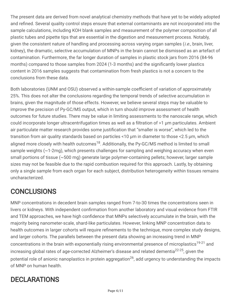
Py-GC/MS has proven to be an informative and reliable method to determine plastics concentrations in liquid and solid tissue samples, with ample assurance of accuracy, quality, and rigor2,3,9,10. Decedent liver and kidney MNP concentrations were similar, with means of 465 and 666 μg/g, respectively, from 2024 samples (Figure 1A). These were higher than previously published data for human placentas (126 μg/g)10, but comparable to testes (329 μg/g)11. Liver samples had significantly higher concentrations in 2024 than in 2016 samples (145 μg/g; p<0.001). The brain samples, all derived from the frontal cortex, revealed substantially higher concentrations than liver or kidney, at 3,057 μg/g in 2016 samples and 4,806 μg/g (0.48%, by weight) in 2024 samples, ranging as high as 8,861 μg/g. Five brain samples from 2016 (highlighted in orange, Figure 1A,,B)B) were analyzed independently by colleagues at Oklahoma State University, and those values were consistent with our findings.
Py-GC/MS는 정확성, 품질 및 엄격성에 대한 충분한 보장을 통해 액체 및 고체 조직 샘플에서 플라스틱 농도를 결정하는 유익하고 신뢰할 수 있는 방법으로 입증되었습니다.2,3,9,10 사망자의 간 및 신장 MNP 농도는 비슷했으며, 2024개 샘플에서 각각 평균 465 및 666 μg/g였습니다(그림 1A). 이는 이전에 발표된 인간 태반 데이터(126 μg/g)10보다 높았지만 고환(329 μg/g)11과 유사했습니다. 간 샘플은 2016년 샘플보다 2024년에 농도가 상당히 높았습니다(145 μg/g; p<0.001). 전두엽 피질에서 추출한 뇌 샘플은 모두 간이나 신장보다 상당히 높은 농도를 보였으며, 2016년 샘플에서는 3,057 μg/g, 2024년 샘플에서는 4,806 μg/g(0.48%, 중량 기준)으로, 최대 8,861 μg/g까지 나타났습니다. 2016년의 5개 뇌 샘플(주황색으로 강조 표시, 그림 1A,,B)B)은 오클라호마 주립 대학의 동료들이 독립적으로 분석했으며, 그 값은 우리의 연구 결과와 일치했습니다.
|
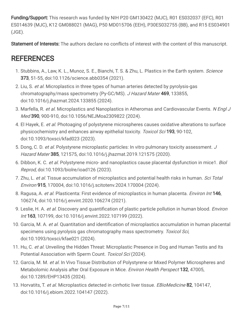
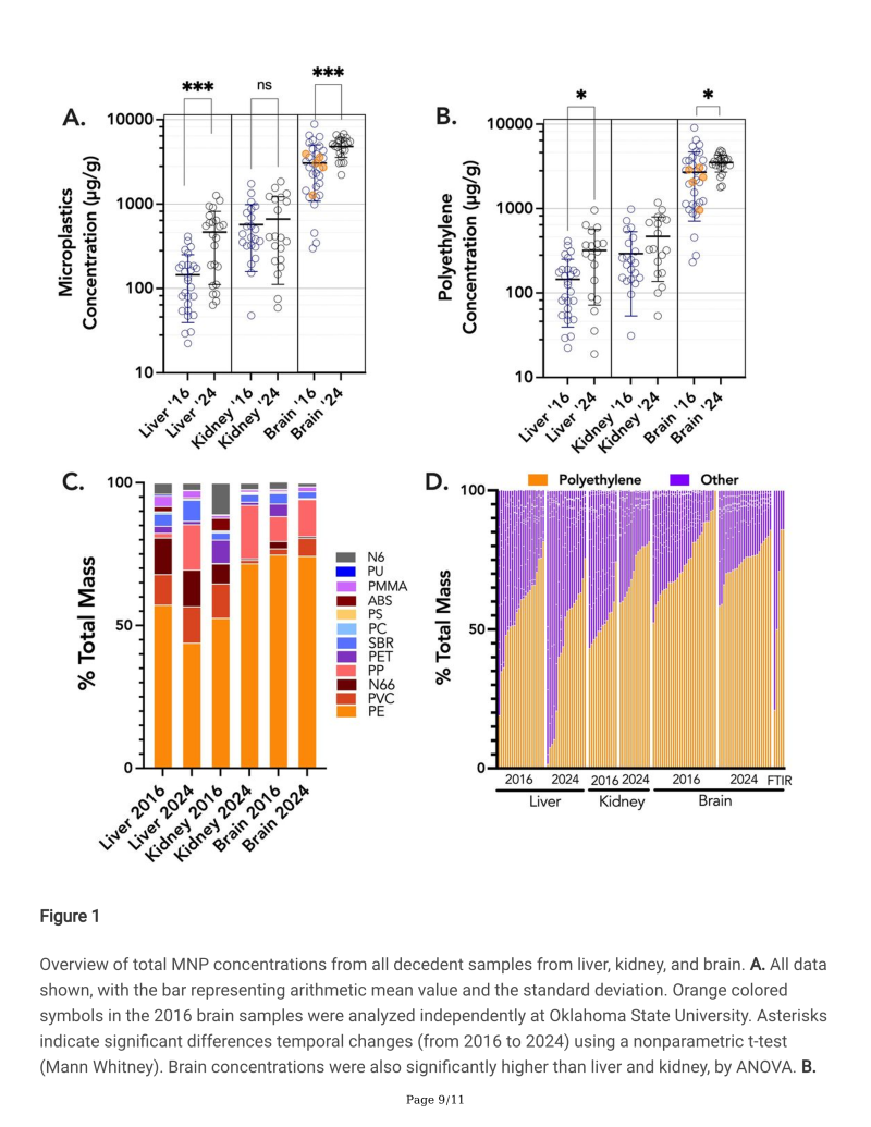
Overview of total MNP concentrations from all decedent samples from liver, kidney, and brain. A. All data shown, with the bar representing arithmetic mean value and the standard deviation. Orange colored symbols in the 2016 brain samples were analyzed independently at Oklahoma State University. Asterisks indicate significant differences temporal changes (from 2016 to 2024) using a nonparametric t-test (Mann Whitney). Brain concentrations were also significantly higher than liver and kidney, by ANOVA. B. Using only polyethylene data, similar trends were noted, although the kidney concentrations did not increase in the 2024 samples. C. Overall distribution of 12 different polymers suggests a greater accumulation of polyethylene in the brain relative to liver or kidney. Polyethylene (PE), Polyvinyl chloride (PVC), Nylon 66 (N66), Styrene-butadiene (SBR), Acrylonitrile Butadiene Styrene (ABS), Polyethylene terephthalate (PET), Nylon 6 (N6), Poly(methyl methacrylate) (PMMA), Polyurethane (PU), Polycarbonate (PC), Polypropylene (PP), Polystyrene (PS). D. Distribution trends for PE across each organ and collection date, including 5 additional samples (on the right) from the 2016 brain collections that were analysed by Attenuated Total Reflectance-Fourier-transform infrared spectroscopy (#FTIR ).
간, 신장 및 뇌의 모든 사망자 샘플에서 얻은 총 MNP 농도 개요.
A. 모든 데이터는 산술 평균 값과 표준 편차를 나타내는 막대와 함께 표시됨. 2016년 뇌 샘플의 주황색 기호는 오클라호마 주립 대학에서 독립적으로 분석되었습니다.
별표는 비모수 t-검정(Mann Whitney)을 사용하여 시간적 변화(2016년에서 2024년)의 유의미한 차이를 나타냅니다.
뇌 농도는 또한 ANOVA에 의해 간과 신장보다 유의미하게 높았습니다.
B. 폴리에틸렌 데이터만 사용하여 유사한 추세가 나타났지만 2024년 샘플에서 신장 농도는 증가하지 않았습니다.
C. 12가지 다른 폴리머의 전체 분포는 간이나 신장에 비해 뇌에 폴리에틸렌이 더 많이 축적됨을 시사합니다. 폴리에틸렌(PE), 폴리염화비닐(PVC), 나일론 66(N66), 스티렌-부타디엔(SBR), 아크릴로니트릴 부타디엔 스티렌(ABS), 폴리에틸렌 테레프탈레이트(PET), 나일론 6(N6), 폴리(메틸 메타크릴레이트)(PMMA), 폴리우레탄(PU), 폴리카보네이트(PC), 폴리프로필렌(PP), 폴리스티렌(PS). D. 2016년 뇌 수집물에서 감쇠 전반사-푸리에 변환 적외선 분광법(FTIR)으로 분석한 5개의 추가 샘플(오른쪽)을 포함하여 각 장기 및 수집 날짜에 따른 PE의 분포 추세.
|
A non-parametric analysis of variance (Kruskal-Wallis) confirmed that MNP concentrations in brains were significantly greater than all other tissues (P<0.0001). Furthermore, from 2016 to 2024, there was a significant increase in MNP concentrations in both livers and brains. The predominant polymer found in all tissues was polyethylene, which independently displayed similarly increasing trends from 2016 to 2024 in the liver and brain (Figure 1B). The proportion of polyethylene in the brain (74%) appeared significantly greater relative to other polymers in comparison to the liver and kidney (44–57%), although kidney samples from 2024 also had increased relative PE (71%; Figure 1C,,D).D). This was also confirmed with ATR-FTIR spectroscopic analysis from 5 brain samples (Figure 1D).
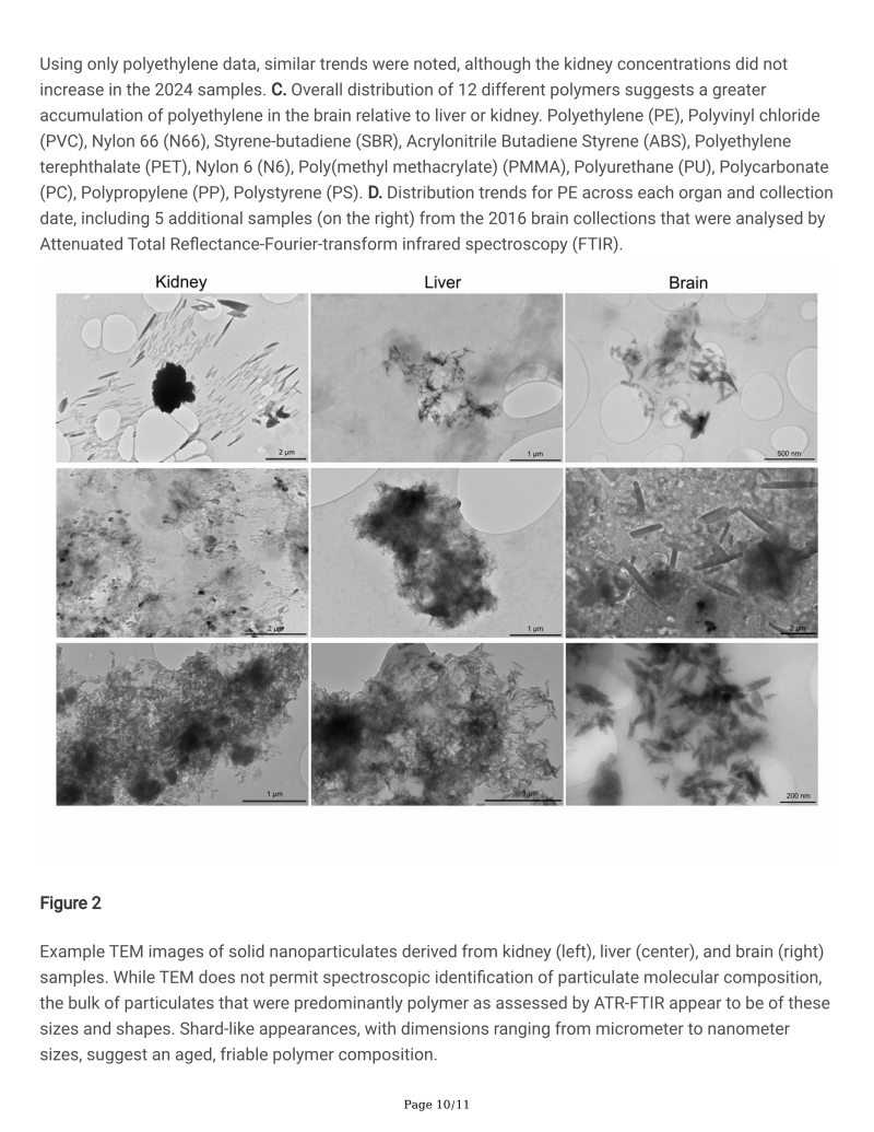
Because we suspected that much of the MNPs measured were actually in the nanoscale range, transmission electron microscopy (TEM) was conducted on the dispersed pellets obtained from kidney, liver, and brain (Figure 2; see methods supplement). While TEM does not provide spectroscopic identification to confirm particulate composition, we observed common shapes and sizes among the numerous samples and tissue types. Notably, there were innumerable particulates with shard-like appearance, often less than 200 nm in length. Currently, MNP uptake and distribution pathways are incompletely understood; this new appreciation of the size and shape aids in our appreciation of potential mechanisms. Importantly, these observations bring into question the relevance of the many recent studies utilizing polystyrene microspheres4,12, as polystyrene was infrequently detected in human tissues and MNPs were rarely spherical.
비모수 분산 분석(Kruskal-Wallis)은 뇌의 MNP 농도가 다른 모든 조직보다 유의하게 높았음을 확인했습니다(P<0.0001). 또한 2016년부터 2024년까지 간과 뇌 모두에서 MNP 농도가 유의하게 증가했습니다.
모든 조직에서 발견된 주요 폴리머는 폴리에틸렌이었으며, 이는 2016년부터 2024년까지 간과 뇌에서 독립적으로 유사한 증가 추세를 보였습니다(그림 1B).
뇌의 폴리에틸렌 비율(74%)은 간과 신장(44–57%)에 비해 다른 폴리머에 비해 유의하게 더 크게 나타났지만, 2024년의 신장 샘플도 상대적 PE가 증가했습니다(71%; 그림 1C,,D).D).
이는 5개의 뇌 샘플에서 ATR-FTIR 분광 분석으로도 확인되었습니다(그림 1D).
측정된 MNP의 대부분이 실제로 나노스케일 범위에 있을 것이라고 의심했기 때문에 신장, 간, 뇌에서 얻은 분산 펠릿에 투과 전자 현미경(TEM)을 수행했습니다(그림 2; 방법 보충 참조). TEM은 입자 구성을 확인하기 위한 분광학적 식별을 제공하지 않지만, 수많은 샘플과 조직 유형에서 공통적인 모양과 크기를 관찰했습니다. 주목할 점은 길이가 200 nm 미만인 파편과 같은 모양의 입자가 무수히 많았습니다.
현재 MNP 흡수 및 분포 경로는 완전히 이해되지 않았습니다. 크기와 모양에 대한 이러한 새로운 이해는 잠재적 메커니즘에 대한 이해에 도움이 됩니다. 중요한 점은 이러한 관찰이 폴리스티렌 미세구체를 활용한 많은 최근 연구의 관련성에 의문을 제기한다는 것입니다.4,12 폴리스티렌은 인간 조직에서 드물게 감지되었고 MNP는 거의 구형이 아니었습니다.
|
Example TEM images of solid nanoparticulates derived from kidney (left), liver (center), and brain (right) samples. While TEM does not permit spectroscopic identification of particulate molecular composition, the bulk of particulates that were predominantly polymer as assessed by ATR-FTIR appear to be of these sizes and shapes. Shard-like appearances, with dimensions ranging from micrometer to nanometer sizes, suggest an aged, friable polymer composition.
신장(왼쪽), 간(중앙), 뇌(오른쪽) 샘플에서 추출한 고체 나노입자의 TEM 이미지 예. TEM은 입자 분자 구성의 분광학적 식별을 허용하지 않지만, ATR-FTIR로 평가한 결과 주로 폴리머인 대부분의 입자는 이러한 크기와 모양을 가진 것으로 보입니다.
마이크로미터에서 나노미터 크기까지 다양한 크기의 파편과 같은 외관은 오래되고 부서지기 쉬운 폴리머 구성을 시사합니다.
|
The concentrations in liver and kidney were not as high (relative to brains) as we would have suspected, as these are “front line” organs for xenobiotic uptake and clearance. That said, the lipophilic nature of plastics may make them easily handled by the liver, which has a major role in uptake and repackaging of dietary triglycerides and cholesterol. A recent study found higher MNP numbers in the cirrhotic liver compared to the healthy liver; whether the microplastics promote disease or are simply accumulating along with intracellular fats has not been elucidated13.
Following this logic, the human brain has the second highest lipid content in the body, with only adipose tissue being higher; brain MNP concentrations are comparable to recently published Py-GC/MS data from carotid plaques, which are also a lipid depot3. Furthermore, the brain receives a high blood flow, approximately 25–30% of the cardiac output, and has a tremendous metabolism. The blood-brain barrier poses a notorious challenge. However, modeling of transfer across cellular membranes suggests the uptake is dependent on the association of particulates with cholesterol and, furthermore, that particles <1μm rapidly traversed the blood-brain barrier within 2h of ingestion in mice14. Longer-term gavage studies similarly found that larger (5 μm) polystyrene microspheres could access the brain and promote metabolomic alterations15. Lastly, clearance rates from the brain are unknown for polymer particulates. The lack of correlation with the decedent age suggests that an equilibrium occurs and may depend on genetic, dietary, and lifestyle factors that ultimately contribute to the wide between-subject variability in MNP concentrations. In zebrafish exposed to constant concentrations, nanoplastics uptake increased to a stable plateau and cleared after exposure16; however, the maximal concentrations were increased proportionately with higher exposure concentrations. While the time course for kinetics is assuredly longer in humans, we postulate that the exponentially increasing environmental concentrations of MNPs1,17 will analogously increase internal maximal concentrations, which is corroborated by our finding that total plastics mass concentration in brains increased over 50% in the past 8 years.
간과 신장의 농도는 우리가 생각했던 것만큼 높지 않았습니다(뇌에 비해). 이들은 이종물질 흡수와 청소의 "최전선" 기관이기 때문입니다. 그렇긴 하지만, 플라스틱의 친유성 때문에 간에서 쉽게 처리할 수 있으며, 간은 식이 트리글리세리드와 콜레스테롤의 흡수와 재포장에 중요한 역할을 합니다. 최근 연구에 따르면 간경변증이 있는 간에서 건강한 간보다 더 높은 MNP 수치가 발견되었습니다. 미세 플라스틱이 질병을 촉진하는지 아니면 단순히 세포 내 지방과 함께 축적되는지는 밝혀지지 않았습니다.13
이 논리에 따르면, 인간의 뇌는 신체에서 두 번째로 지질 함량이 높고 지방 조직만이 더 높습니다.
뇌의 MNP 농도는 지질 저장소이기도 한 경동맥 플라크에서 최근에 발표된 Py-GC/MS 데이터와 비슷합니다.3
게다가 뇌는 심장 출력의 약 25~30%에 해당하는 높은 혈류를 받고 엄청난 신진대사를 합니다.
혈액-뇌 장벽 (#BBB )은 심각한 높은 문제를 제기합니다. 그러나 세포막을 통한 전달 모델링은 흡수가 입자와 콜레스테롤의 연관성에 따라 달라지며, 나아가 1 μm 미만의 입자는 마우스에서 섭취 후 2시간 이내에 혈액-뇌 장벽을 빠르게 통과한다는 것을 시사합니다14.
장기간의 위관 영양 연구에서도 마찬가지로 더 큰(5 μm) 폴리스티렌 미세구가 뇌에 접근하여 대사체 변화를 촉진할 수 있다는 것을 발견했습니다15. 마지막으로, 폴리머 입자의 경우 뇌에서의 제거율은 알려져 있지 않습니다.
사망자 연령과의 상관 관계가 없다는 것은 평형이 발생하고 궁극적으로 MNP 농도의 피험자 간 변동성에 기여하는 유전적, 식이 및 생활 방식 요인에 따라 달라질 수 있음을 시사합니다. 일정한 농도에 노출된 다니오에서 나노플라스틱 흡수는 안정적인 평탄부로 증가한 후 노출 후 제거되었습니다16.
그러나 최대 농도는 노출 농도가 높아질수록 비례적으로 증가했습니다. 인간의 경우 동역학에 대한 시간 경과가 확실히 더 길지만, 우리는 MNPs1,17의 환경 농도가 기하급수적으로 증가함에 따라 내부의 최대 농도도 유사하게 증가할 것이라고 가정합니다.
이는 지난 8년 동안 뇌의 총 플라스틱 농도가 50% 이상 증가했다는 우리의 연구 결과에 의해 입증됩니다.
LIMITATIONS 제한 사항
The present data are derived from novel analytical chemistry methods that have yet to be widely adopted and refined. Several quality control steps ensure that external contaminants are not incorporated into the sample calculations, including KOH blank samples and measurement of the polymer composition of all plastic tubes and pipette tips that are essential in the digestion and measurement process. Notably, given the consistent nature of handling and processing across varying organ samples (i.e., brain, liver, kidney), the dramatic, selective accumulation of MNPs in the brain cannot be dismissed as an artefact of contamination. Furthermore, the far longer duration of samples in plastic stock jars from 2016 (84–96 months) compared to those samples from 2024 (1–3 months) and the significantly lower plastics content in 2016 samples suggests that contamination from fresh plastics is not a concern to the conclusions from these data.
Both laboratories (UNM and OSU) observed a within-sample coefficient of variation of approximately 25%. This does not alter the conclusions regarding the temporal trends of selective accumulation in brains, given the magnitude of those effects. However, we believe several steps may be valuable to improve the precision of Py-GC/MS output, which in turn should improve assessment of health outcomes for future studies. There may be value in limiting assessments to the nanoscale range, which could incorporate longer ultracentrifugation times as well as a filtration of >1 μm particulates. Ambient air particulate matter research provides some justification that “smaller is worse”, which led to the transition from air quality standards based on particles <10 μm in diameter to those <2.5 μm, which aligned more closely with health outcomes18. Additionally, the Py-GC/MS method is limited to small sample weights (~1–2mg), which presents challenges for sampling and weighing accuracy when even small portions of tissue (~500 mg) generate large polymer-containing pellets; however, larger sample sizes may not be feasible due to the rapid combustion required for this approach. Lastly, by obtaining only a single sample from each organ for each subject, distribution heterogeneity within tissues remains uncharacterized.
현재 데이터는 아직 널리 채택되고 개선되지 않은 새로운 분석 화학 방법에서 파생되었습니다.
여러 품질 관리 단계를 통해 KOH 블랭크 샘플과 소화 및 측정 프로세스에 필수적인 모든 플라스틱 튜브 및 피펫 팁의 폴리머 구성 측정을 포함하여 외부 오염 물질이 샘플 계산에 포함되지 않도록 합니다.
특히 다양한 장기 샘플(예: 뇌, 간, 신장)에서 일관된 취급 및 처리 특성을 감안할 때 뇌에서 MNP의 극적이고 선택적 축적을 오염의 인공물로 기각할 수 없습니다.
또한 2016년(84~96개월)의 플라스틱 스톡 항아리 샘플 기간이 2024년(1~3개월)의 샘플에 비해 훨씬 길었고 2016년 샘플의 플라스틱 함량이 상당히 낮았기 때문에 신선한 플라스틱으로 인한 오염이 이러한 데이터의 결론에 문제가 되지 않음을 시사합니다.
두 실험실(UNM 및 OSU) 모두 약 25%의 샘플 내 변동 계수를 관찰했습니다.
이는 해당 효과의 규모를 감안할 때 뇌의 선택적 축적의 시간적 추세에 대한 결론을 바꾸지 않습니다.
그러나 Py-GC/MS 출력의 정밀도를 개선하는 데 몇 가지 단계가 중요할 수 있다고 생각하며, 이는 향후 연구를 위한 건강 결과 평가를 개선해야 합니다.
평가를 나노스케일 범위로 제한하는 데 가치가 있을 수 있으며, 여기에는 더 긴 초원심분리 시간과 >1 μm 입자의 여과가 포함될 수 있습니다.
주변 공기 입자 물질 연구는 "작을수록 더 나쁘다"는 것을 어느 정도 정당화하는데, 이는 직경이 <10 μm인 입자를 기반으로 하는 대기 질 기준에서 건강 결과와 더 긴밀하게 일치하는 <2.5 μm 입자로 전환하는 데 도움이 되었습니다.18
또한 Py-GC/MS 방법은 작은 샘플 무게(~1–2 mg)로 제한되어 조직의 작은 부분(~500 mg)에서도 큰 폴리머가 포함된 펠릿이 생성될 때 샘플링 및 무게 측정 정확도에 어려움이 있습니다.
그러나 이 접근 방식에 필요한 빠른 연소로 인해 더 큰 샘플 크기는 실행 불가능할 수 있습니다.
마지막으로, 각 피험자의 각 장기에서 단 하나의 샘플만 얻었기 때문에 조직 내 분포의 이질성은 여전히 특성화되지 않았습니다.
CONCLUSIONS 결론
MNP concentrations in decedent brain samples ranged from 7-to-30 times the concentrations seen in livers or kidneys. With independent confirmation from another laboratory and visual evidence from FTIR and TEM approaches, we have high confidence that MNPs selectively accumulate in the brain, with the majority being nanometer-scale, shard-like particulates. However, linking MNP concentration data to health outcomes in larger cohorts will require refinements to the technique, more complex study designs, and larger cohorts. The parallels between the present data showing an increasing trend in MNP concentrations in the brain with exponentially rising environmental presence of microplastics19–21 and increasing global rates of age-corrected Alzheimer’s disease and related dementia22–25, given the potential role of anionic nanoplastics in protein aggregation26, add urgency to understanding the impacts of MNP on human health.
사망자 뇌 샘플의 MNP 농도는 간이나 신장에서 관찰된 농도의 7~30배에 달했습니다.
다른 실험실의 독립적인 확인과 FTIR 및 TEM 접근법의 시각적 증거를 통해 MNP가 뇌에 선택적으로 축적되며 대부분이 나노미터 크기의 파편과 같은 입자라는 확신이 큽니다. 그러나 MNP 농도 데이터를 대규모 코호트의 건강 결과에 연결하려면 기술 개선, 더 복잡한 연구 설계 및 대규모 코호트가 필요합니다.
뇌의 MNP 농도가 기하급수적으로 증가하는 환경적 미세플라스틱19–21과 연령 교정 알츠하이머병 및 관련 치매22–25의 전 세계적 증가율을 보여주는 현재 데이터 간의 유사점은 단백질 응집26에서 음이온 나노플라스틱의 잠재적 역할을 감안할 때 MNP가 인간 건강에 미치는 영향을 이해하는 것이 시급하다는 것을 더합니다.
Funding/Support:
This research was funded by NIH P20 GM130422 (MJC), R01 ES032037 (EFC), R01 ES014639 (MJC), K12 GM088021 (MAG), P50 MD015706 (EEH), P30ES032755 (BB), and R15 ES034901 (JGE).
Footnotes
Statement of Interests: The authors declare no conflicts of interest with the content of this manuscript.


https://www.ncbi.nlm.nih.gov/pmc/articles/PMC11100893/
Bioaccumulation of Microplastics in Decedent Human Brains Assessed by Pyrolysis Gas Chromatography-Mass Spectrometry
Rising global concentrations of environmental micro- and nanoplastics (MNPs) drive concerns for human exposure and health outcomes. Applying pyrolysis gas chromatography-mass spectrometry (Py-GC/MS) methods to isolate and quantify MNPs from human samples,
www.ncbi.nlm.nih.gov
******************************************************************************************************
#미세플라스틱 #시험분석 기술서비스 #시험표준개발 #국제공인시험기관 #KOLAS
#시험/분석 #환경 #수질 #토양 #대기 #폐기물 #타이어 #슬러지
#식품 #음료 #벌꿀 #주류 #소금 #어패류 #해조류 #미세플라스틱분석 #미세플라스틱시험
#세탁폐수 #미세섬유 #세탁망 #필터 #화장품 #치약 #생활화학제품 #세탁기필터
#식품용기 #티백 #젖병 #종이컵 #NSF 평가 #젖병소독기 #정수기미세플라스틱시험
#표준개발 ISO/TC61/SC14, TC38, TC147/SC2&SC6 (Microplastics) Korean Delegate
IEC/TC 111/WG 3 & JWG 14 Co-Convenor
IEC 62321-3-2(#Halogen ),-10 (#PAHs ), -13(#BPA ), -14(#SCCP/MCCP ) Project leader
- 분석장비: #TEDGCMS, #microFT-IR #microRaman, #ICPMS #XRF #Combustion-IC 등
#PAHs분석 #SCCP/MCCP분석 #Halogen분석 #PAHs #SCCP #MCCP #halogen #PFAS
#생체시료분석 #생체미세플라스틱분석 #혈액미세플라스틱분석 #인체미세플라스틱분석
#생체시료시험 #생체미세플라스틱시험 #혈액미세플라스틱시험 #혈액미세플라스틱검사
#미세플라스틱RM #미세플라스틱표준시료 #미세플라스틱표준물질 #태블릭RM
#ISO/IEC17025 ( #국제공인시험기관 , #KOLAS ) 인정 연구소
******************************************************************************************************